

2D Echo
A 2D echocardiogram, also known as 2D Echo, is a non-invasive diagnostic test that uses ultrasound waves to create detailed images of the heart. This imaging technique allows healthcare professionals to visualize the structure and function of the heart in real time. Here are key aspects of a 2D echocardiogram:
How 2D Echo Works:
- Ultrasound Waves:
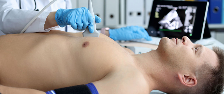
- The test involves the use of high-frequency sound waves (ultrasound) that are directed toward the heart from a small hand-held device called a transducer.
- Reflection of Sound Waves:
- As the sound waves encounter different structures in the heart, such as chambers, valves, and blood vessels, they reflect back to the transducer.
- Image Formation:
- The reflected waves are then converted into visual images on a computer monitor, creating a real-time moving picture of the heart.
Components of a 2D Echocardiogram:
- Two-Dimensional Images:
- Provides a detailed view of the heart’s chambers, valves, and surrounding structures in two-dimensional images.
- M-Mode Imaging:
- Motion-mode imaging displays a one-dimensional slice of the heart over time, allowing for precise measurements of cardiac structures and movements.
- Doppler Imaging:
- Doppler ultrasound assesses blood flow through the heart and blood vessels. It can help detect abnormalities such as regurgitation, stenosis, or shunting.
Uses of 2D Echo:
- Structural Assessment:
- Evaluates the size, shape, and thickness of the heart chambers, as well as the condition of the heart valves.
- Function and Motion:
- Assesses the movement and contraction of the heart muscle (myocardium) to determine its pumping function.
- Valve Function:
- Examines the opening and closing of heart valves to identify abnormalities such as regurgitation (leakage) or stenosis (narrowing).
- Congenital Heart Conditions:
- Detects and evaluates congenital heart defects in both adults and children.
- Cardiac Tumors:
- Helps identify tumors or masses within the heart.
- Pericardial Diseases:
- Assesses the condition of the pericardium (the sac around the heart) for abnormalities or fluid accumulation.
- Cardiac Function in Disease States:
- Provides valuable information in conditions such as heart failure, cardiomyopathy, and ischemic heart disease.
- Guidance for Procedures:
- May be used to guide certain cardiac procedures, such as pericardiocentesis or valve repair.
Types of 2D Echocardiograms:
- Transthoracic Echocardiogram (TTE):
- The most common type, where the transducer is placed on the chest wall to obtain images through the chest.
- Transesophageal Echocardiogram (TEE):
- Involves inserting a specialized transducer into the esophagus to obtain clearer images of the heart structures, particularly in patients where TTE may not provide adequate views.
- Stress Echocardiogram:
- Performed during exercise or pharmacological stress to assess heart function under challenging conditions.
2D Echo is a valuable tool for diagnosing and monitoring various cardiovascular conditions. The results of the test aid healthcare professionals in formulating treatment plans and assessing the overall health of the heart. It is commonly used in cardiology practice and is a routine part of cardiac evaluations.
